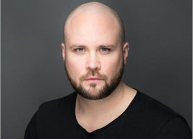
Animal models predominate the field of drug testing and discovery, despite a growing body of evidence suggesting that such models are often poor predictors of human reactions. As a postdoctoral researcher at the University of Lausanne in Switzerland, Dr. David Pamies is working on something which might help to address these shortcomings: a human-induced pluripotent stem cell-derived 3D platform, and the development of a brain organoid—an in vitro model representing the human brain.
He and his team are using the model to predict the toxicity of many different chemicals that we encounter on a daily basis. In addition to providing a cheaper and more efficient way of studying this than the industry standard of mouse models, this model is allowing Dr. Pamies to study the effectiveness of pharmaceuticals such as temozolomide in glioblastoma, a type of brain cancer also referred to as glioblastoma multiforme (GBM).
He explains how they transplant and incorporate glioblastoma tumors from humans into the model, what they’ve learned so far in doing so, and the current issues they’re working on. He also discusses the nature of his most recent research and where it’s headed in the future.
Tune in to hear the full conversation.
Here are different publications related to Organoids and Human Brain:
Organanoids, 3D cultures.
Glioblastoma model:
Organoids vascularization:
Richard Jacobs: Hello, this is Richard Jacobs with the future tech and future tech health podcast. I have David Pamies. He’s on the faculty of biology and medicine, the University of Lausanne in Switzerland, used to be at Johns Hopkins, and we’re going to be talking about a human induced pluripotent STEM cell-derived 3d platform. So, David, thank you for coming.
David Pamies: No, thank you for having me. It’s a pleasure.
Richard Jacobs: For people that don’t know what is induced pluripotent STEM cells?
David Pamies: So pluripotent STEM cells are basically when you have adversarial and then you can reprogram the cells to become, again as STEM cells. So that allows you to get STEM cells from different patients without going through taking the cells from the embryos. That was a big ethical problem before. So you can introduce certain factors inside the somatic cells and they become again STEM cells. So that’s basically induced pluripotent.
Richard Jacobs: Right. What cells are you trying to recreate in your particular experiments?
David Pamies: So what we are trying to develop is in vitro models that represent a human brain. So basically we are trying to do it in a 3D structure. So basically we generate like a small tissue that has different neuronal cell types as to size and illegal in the size, the different types of cells that we will find in the brain. So we differentiate them all together so they are interconnected and they assembly a little bit or mimic a little bit what we will find in the brain tissue.
Richard Jacobs: The brain is incredibly complicated. So how much of the brain function that you’re trying to approximate with your spheroid?
David Pamies: That’s a good question. So in our case, the application we use this for is toxicology. So we need to find a way that we can find something that is complex enough to kind of reproducing some functionality of the brain. But that is not super complex because when you get like a really complex model, then it’s really difficult to have a reproducible system. So in toxicology, we want to have a model that every time we produce these tissue is the same. So then when we introduce chemicals and toxicants or drugs we don’t have the viability of the older model. In our case, what we have is let’s say the basic tissue you need. So we have the neurons that are connected between them and they have a spontaneous electrical activity. We have the illegal intercise rapping with the male in the exons and as to say, support in neurons. But we didn’t have these like the different regions of the brain. There’s obviously some parts of the brain that have specific types of neurons or things like that. So we have something that is kind of general so that they reproduce a little bit what will be the general structure of the brain but it doesn’t have the particularly of each different region in the brain.
Richard Jacobs: Yeah. Well, sure, that’d be really impossible to recreate. Are you assuming in terms of toxicology that the substances you’re testing have made it past the blood-brain barrier?
David Pamies: Yes. So we also have incorporated in our model blood-brain barrier systems. So we can check if the chemicals with passive blood and barrier or not before testing in our system. But also there is by chemical properties. So the drug, sometimes you already know it, this is something that is going to happen most likely or not. But you can also test this in vitro to check if the chemicals will cross the blood-brain barrier or not.
Richard Jacobs: And where do you get these cells from? Do they start out as skin cells? I mean, what do they start out as before you bring them back to pluripotency?
David Pamies: Normally we get them from donors. We take from their skin and then from the skin we rep around them to become STEM cells. And at the moment we have several lines that are coming from healthy donors, but also from some disease. So we have, for example, Parkinson’s disease. We have some autistic lines from Autism and things like that. When we get the glioblastoma cause we also use the mogul for a dragger screening in glioblastoma research. Then the glioblastoma is obtained directly from the patient. So when the patients die, then they remove the glioblastoma and then we incorporate these cells into these brain spheres.
Richard Jacobs: So have you seen that certain candidate conditions do translate to the cells themselves? They’re preserved even through pluripotency and certain ones are not?
David Pamies: From my experience, I haven’t seen much difference. There are so many studies showing the differences between diseases. In my hands, I haven’t seen that, but there are some studies, for example, some schizophrenic patients have a lower alienation in the exons and things like that. However, sometimes it’s really difficult to unknown to know if it’s because of the disease or because of the reprogramming. So you have to be really sure that the cells, it could be also by the variability between individuals and not because of the disease. So you need to have several lines from patients that you have to have several lines from healthy patients and then you have to derive in the same ways so that because there are different ways to wrap around the cells and differentiate them. So you had to make a lot of controls that have a lot of lines to make sure that the differences are because of the disease and not because of the variation of their process. But there is some interesting research already published showing the differences in some diseases.
Richard Jacobs: When does brain toxicity tend to occur when someone takes a drug? Is it very quickly or does it happen later on at the drug has been metabolized by the liver and other organs?
David Pamies: Well, what we are doing is a little bit different. What we are trying to do is to predict the toxicities that will happen in humans. So at the moment, the current guidelines are important mainly. So people use rats or mice to do different tests to see if that chemical is a teratogenic or produce high irritation or things that that so what we’re trying to develop a test that is a human-based test and also in vitro. So is faster and cheaper because at the moment the problem is that we have so many chemicals without knowing what are the toxic effects and the guidelines are super slow and super expensive. So it’s really difficult to test all the chemicals. So we are trying to find something that is really fast in order to predict these chemicals. We are not in the pharma kinetics. So we are not checking how the drags are going through the body and so on. We are more trying to identify which chemical produces some effects in the brain. Chemicals that are exposed in the environment, for example, the flame retardants that we have in the tables to avoid that they start a fire. And this kind of chemicals we are exposed to all the time and we don’t know what effects they have in our brain. So what we are trying to develop a test that is cheap and fast enough to be able to test many chemicals and whether they’re mimic, you know, human physiology enabled to predict better than the animals because at the moment animals have a really bad revision or also in humans. And recently we have also tried to study in the drug development field, but mostly focused on toxicology. Also how different drugs will affect glioblastoma tumors. But also kind of in the same way. So we are now working on how the chemical goes through the body and is metabolite, is mostly to see which drugs are more efficient to kill the glioblastoma tumors without killing the healthy cells. So this is more or less
Richard Jacobs: But the issue may be what if you test all these drugs and they don’t look clean in toxicity but in vivo, let’s say they’re metabolized by the liver and processed by different parts of the body and then they’re toxic. We wouldn’t know that without hooking up a bunch of other organoids from different parts of the body.
David Pamies: You are completely right. So what people are doing now is they are trying to develop esilico models. So they are trying to do direct the studies into how the drugs are metabolized and so on. And then tried to model by income by esilico. So they are doing computer science. They tried to model a model that could predict how the chemicals are going to be distributed in the body, how they are going to be metabolized and so on. So then they try to compare this in vivo and try to see if the models work and things like that. And also there are some people trying now to link this with a chemical structure so they can predict, for example, if you know that these ten chemicals have a similar chemical structure and they will be metabolized in a certain way or distributed in their body in a certain way, then how much you can predict the chemicals that also have a similar chemical structure. And that’s how people are trying to do this. However, yeah, if it’s intense of a drug until you go to clinical trials and things like that is really hard to know. I want 100% of what is going to happen with the track. By the way, you want to at least try to have the safeties drug going to clinical trials. And it’s a big problem at the moment because it’s the same way that with the animals, so if you check the statistics that how many drugs that are developed going to the market. Only like 10% of the drugs they will up going to the market. And mostly they fail in the clinical trials. And this is because the main modeling we have at the moment and also with the in vitro system, you are trying to develop something in a rat model that obviously is not a human and then you use different concentrations per kilogram and things like that. And then when you moved to a human, then most of the problems with the drug are relative with efficacy or with safety. So the drugs don’t behave as you were expecting and also they are more toxic than they were expected in humans. And that’s why all the drugs fail. So this is a big issue at the moment to try to figure it out, how we can solve this problem and how we can make drug candidates more efficient, the selection of the candidates more efficient so we don’t waste so much time because clinical trials are really expensive. So you have to be 100% sure that you are going to stay clinical trials because people don’t have money to do many of those.
Richard Jacobs: But if clinical trials are so expensive, why don’t the various organoid creators get together and cross-license the organoids so that they can get a much better approximation of what really goes on. Or like, let’s say you’re going to test 500 compounds this year. If you don’t create the liver organoids yourself, why not partner with a company that does and send them all the chemicals and partner with them on the results, Hey, when you guys run it through your liver chip are you getting the same thing out as we are getting?
David Pamies: Yeah, I totally agree with you. I think there are people trying to connect different organoids together into like that’s what a few years back I was in this huge project in US that was funded by FDA and DARPA and NIH trying to do the human and a chip. So they will have all these organoids together into the chip and there are some publications showing the connection of a few organoids. Like for example liver, kidney but to connect all the organoids at the moment is not so easy because you need to solve the problem of having the same immedia for all the cells. Also how can have similar States of differentiation in all the cells because cells may be in the definitive stages, they see even some sets that will never be able to complete a few in my tray. So it’s not so easy to do this, but I agree that if we share more the information it could be easier to reach a good point. However, pharma doesn’t want to normally share any information about anything that could be profit from them. It’s already high in like public science because publications so you put in all the pressure that you want to be the first and things like that. But if you moved to pharma companies it’s even worse because you know, they know that if they have a really good candidate, it could be a lot of money. So if you want to know, you know how this is metabolizing another organoid but you don’t have it in house, you have to share the information with someone else and this type of thing. It gets complicated. And also I think pharma is all these organoids and organ to achieve things are really new. I think pharma is still like a little bit like seeing what happened and before going into this type of research and we know because we have some contact with some pharma industry and they are in Switzerland. They are really interested, but they obviously don’t have yet in the house any of those systems. So they are talking with us to see how they can use the models and how they can incorporate these pipelines or the initial driver screening and things like that. But as it is quite new I think pharma still a little bit not really into the game yet.
Richard Jacobs: Who first came up with the idea of an organoid and when? Do you know?
David Pamies: Well 3D models so it depends who are you consider going to because also the terminology side changing in the last years. But it has been already known for a long time 3D cultures and 3D of course that already resemble a little bit of functional bio organ. And these have been really from really long time. In the case of the brain in 2013, there was a Lancaster paper that was, I think was a nature paper and they were the first time to show that they can differentiate the brain with a little bit of the structure of the initial development of the brain. So they have different compartments of the brain with different sections. Now you could distinguish between forebrain, midbrain and these type of things. And this was in 2013, but there are all the organs, like the liver, organoids and things like that. That may be a little bit earlier than that, but I would say maybe four for the new organoids, I would say maybe 14 years or maybe 10 years. But there have been already people working on rats organoids, rat’s 3D models from a really, really long time ago.
Richard Jacobs: In making the organoid, you’re just making like a spheroid that, I mean are you seeing that the cells are moving around and rearranging and creating their own morphologies as you differentiate them or like what do they do? Is any interesting behavior there?
David Pamies: There is definitely some. They did make me a little bit depressed that we could find in the development of the rain. For example, you could see how the different cell populations will differentiate in different stages. So for example, you will see first the differentiation of neurons and astrocytes and then the oligodendrocytes come in later on. And then the oligodendrocytes after a while they’re starting producing the mailing that will wrap the exons. These types of things you can see them. Also, you can see, for example, the high proliferation of oligodendrocytes at the beginning of they start to differentiate. They are selecting only the oligodendrocytes as functional and the other ones are dying over the differentiation process and this is something that also happened into the normal development of the brain. So you can see these types of things. It depends on how complex is the model. If you go for like several organoids, you can see also how the extent cells migrate and then differentiate over the migration and the different stages of differentiation through the migration and these types of things. I only talk about the brain because this is my field, but I’m pretty sure you can see also in other organoids. How is mimic kind of the differentiation that happened in the normal and Vivo situations
Richard Jacobs: Are you using scaffolding or how do you make sure that the organoids form in some kind of sensible way?
David Pamies: In our case, we are using bioreactors. So basically the cells are floating and it has a certain movement to avoid that they grow in size. Because in our case, we want to keep them smaller than 400 microns because when they, for example, the organoids that we were talking that West Bobby’s by Lancaster, they grow really big and they have different sizes. However, they starting getting aquatic centers in the middle and that in toxicology or drug screening is not possible to use. Because then you don’t know if the toxicant that you’re put in is the one causing these necrotic centers or not. So we are trying to keep this to the nearest sphere. So our brain spheres say smaller than 400 microns, so they don’t get an aquatic center. So we use this a spinning moment to keep them really homogeneous in size and in shape. So that’s how we culture the model.
Richard Jacobs: So the organoids form into like a spheroid shape of what dimension?
David Pamies: It’s like a sphere and it’s around 350 microns. When we introduced the tumors so, as I say, sometimes we use these for a drag a screening for a glioblastoma. What we do is incorporate a few cells of the tumor inside the sphere and then over the weeks the tumor is growing and start interacting with the cells in this are running. And then in this case, sometimes you can see that the brain spheres with the tumor grow bigger than 350 micros, but is because of the growth of the tumor.
Richard Jacobs: Interesting. As you’re modeling these tumors as well, and then testing drugs on the organoid with the tumor in it.
David Pamies: Yeah, I think we are the first doing these. Normally in glioblastoma tumor feel the people have been using the two more derive cells from patients. And then growth in 3D. However, they are growing normally only the tumor cells and they have been testing different chemicals in these tumor cells. However, we know that the tumor glioblastoma interacts with the cells in the surrounding and there is a crosstalk between the healthy cells and the tumor for the progression and the survival of the tumor. So I think we wanted to be sure that we have the tumor into a microenvironment of the tumor. So we can also see the interactions between the tumor on the surface and the surrounds. But also this is really interesting when we do the drug screening because normally drug screening is based on the tumor. So you will see the effects of the drugs into the tumor, but you don’t know if the drug is also going to affect the healthy tissue because some of the chemotherapies also are toxic, they have some toxic effects in the healthy brain. So with this system, you can check the tumor if the tumor is affected by the drug. But also if the healthy cells are affected by the drug. And we have incorporated this in a high school food platform. So we can measure hundreds of drugs at the same time. And now we’re trying to do a bigger test with more than a hundred chemicals into the drugs in the glioblastoma that we have.
Richard Jacobs: And as the tumor act versus the regular cells, do you see differences in behavior and so what do you notice about them that’s different?
David Pamies: Yeah, well, first the tumor is growing faster. And normally when we have the brain spheres when cells differentiate, normally they don’t proliferate a lot. For example, neurons will not proliferate after they are mature. So at some point they brain spheres stop growing. They are increasing the connections between the cells and stuff like that, however, the tumor still growing and you could see some markers that are for proliferation that are really present in the tumor, well in the healthy tissue you don’t see it. And what we are trying to do now is we are going to try to study why are the molecules that the tumor is to creating to the healthy cells and how the tumor is changing the healthy cells around the tumor. That’s what we are trying to focus on now. But definitely the tumor with the healthy cells they have completely different behavior.
Richard Jacobs: Is there any immune system that normally would be in the brain? And are you able to mimic that in the organoid at all?
David Pamies: No, actually immune cells are really important for development for glioblastoma development. And in fact, the microglia that is the main immune cell in the brain is really interacting with the tumor and the microglia migrate into the tumor. And they are the main key players in the model. Because of the microglia, the right from differences not from the initial sources of what we use. The microglia are derived from a different lineage. So what we are doing now is in collaboration with the Sally Olli Nokso university. We are deriving microglia from IPS’s and we are incorporating also the microglia into the sphere. So we have the brains spheres with microglia on and we already have VC in house and we are trying to activate the microglia with a different stimuli with inflammation cytokines or things like that. And what we are trying to do now is also to incorporate the tumor and see how different, for example, we are trying different drugs like for example doxorubicin and we want to see if the sensity of the drugs changed when we had the microglia into our model. So that’s something that we are working on that because we know that microglia is really important for glioblastoma development. So we are trying now to solve that issue and hopefully, we’ll get some interesting results in the next year.
Richard Jacobs: This is a weird question, but could you ever grow an organoid just from cancer cells? Do they differentiate in form and structures or they just seem to just go on a dividing rampage and they don’t bother to differentiate it or anything
David Pamies: Actually, in the glioblastoma field, when they take a glioblastoma from a patient and they do some cultures derive from these glioblastomas, in many cases they call them organoids. So how different cell types into the glioblastoma and also you can see certain kinds of structure. So it is not like the glioblastoma is simulating any organ, but they had the allness structure into the glioblastoma tumor per se. So they started getting aquatic centers and that will help to secrete some growth factors that will bring the vasculature or things like that and you can see how sales are distributed differently into the tumor. So there are many people already call them organoids to these tumors per se because they have a kind of a specific structure.
Richard Jacobs: Yeah. I just wonder, I mean, could you make, so you literally could have a brain organoid that is entirely cancer cells and would it look anything like an organoid of healthy cells?
David Pamies: In my opinion it means that they simulating something that is happening. So you can have an organoid that is simulating what is the brain and you can have an organoid that’s simulating the tumor. They are completely different. One is simulating like me trying to represent like healthy tissue. And the other one is trying to represent more like a tumor tissue. Other people say that they are going to TP cultures and microfiche geological systems. And at the moment it is a little bit weird all these definitions and also people in cancer have been calling these organoids for a long time and now we use the word for other things and it’s kind of confusing.
Richard Jacobs: It makes sense. So any really interesting things that you’ve discovered recently from your work on these organoids that you can talk about? Anything that jumps out of the shoe that was really surprising?
David Pamies: Well, I think the most interesting thing that we have been like I say, we are more into the testing field. So something that we have been studying lately has been an antidepressant drug that pregnant women normally could take it and we have been studying the effects of this drug in the brain development and what we have developed a test that has different endpoints that measure different key events during the brain development. Like for example, geneses, male information or legal intercise maturation. So several things into the same model. So we have to face the drug with a blood concentration that pregnant women will have. And we have this in our system and we have been able to see some effects in the brain development into our test indicating that this drug might have some effects on the development of their brain when the pregnant women are taken. And that could be very interesting to see. If that is the case then it would be allowed in the government. So that we can restrict the use of these drugs.
Richard Jacobs: So some of the organoids are they made from male lineages or female lineages? And is there any difference when you culture them?
David Pamies: So we have serial lines we have from males and from the female. We haven’t seen differences in terms of differentiation. At least the lines that we have. We haven’t done any study comparing a female and a male at the moment, but we don’t see differences in terms of differentiation but there might be some differences in terms of some proteins. But as we don’t have the hormones into the differentiation process and almost in my view really important to see the differences between sex females and males. We haven’t seen yet anything at the moment. I don’t know because we don’t work in that field a lot, but I know that many people now are trying to see the differences between men and women lines. There is obviously a difference between individuals. So there’s some polymorphism that might make some lines more sensitive to certain chemicals than others. That’s why it’s important to study not only one line, and when you test that chemical you want to have a serial line, so you at least have a little bit of heterogeneity in terms of polymorphous and things like that.
Richard Jacobs: One thing I haven’t asked, I forgot, how the organoids get food, nutrients, and oxygen? How do they respirate and how do they get food?
David Pamies: In the media, it already has the nutrients that they need. They basically absorbed the nutrients from the media. So it will be like the extracellular will provide the nutrients, the oxygen, also as we don’t have the vascular tubes. And at the moment there is no much abundance in trying to put vascular to in the organoids. They also have to get the oxygen through the basically taking from the media without any vascular tube that brings the oxygen. And that’s why when you get that really big organoid, you start seeing how the cells in the middle are dying because they oxygen cannot fuse into the centers of these organoids. And I know there are already some publications showing that they are trying to generate also I’ve asked you that to or into the organoids. So then the oxygen will be able to reach all cells into the organoids. But at the moment you can only see like there are no complete vessels. And it’s not completely functional yet. But I know there are some people trying to work on this to be able to reach the oxygen in on the cells. Also, many people use bioreactors or shakings. So the cells are in movement and you can have a higher interchange of oxygen between the media and the incubator. But that’s all the limited, you cannot get really big tissue with a vascular tube because you know, not all the cells will be able to get the nutrients on the oxygen.
Richard Jacobs: Okay. Got it. Well, very good. So what’s the best way for people to find out more about your work and maybe contact you and interact?
David Pamies: If they want to contact me, they can look for me on the website of the university or the department of physiology at Lausanne University. If there are scientists interested, they kind of look for my publications and the corresponding outer in most of the last publications. So my email is there. So they can contact me.
Richard Jacobs: Okay. That’s great. Well, David, thank you for coming. It’s been a very good call.
David Pamies: Thank you very much for having me.
Podcast: Play in new window | Download | Embed


